
Keypoints
What is known
– Current evidence is insufficient to recommend routine therapeutic hypothermia in mild neonatal hypoxic-ischemic encephalopathy.
– Brain injury and neurodevelopmental impairments occur in neonates with mild hypoxic-ischemic encephalopathy.
– Treatment rates with therapeutic hypothermia in newborns with mild hypoxic-ischemic encephalopathy are increasing.
What is added
– This is the first report describing the use of therapeutic hypothermia in mild hypoxic-ischemic encephalopathy in Portugal.
– The proportion of neonates with mild hypoxic-ischemic encephalopathy receiving therapeutic hypothermia is lower than that reported internationally.
– The profile of treated patients did not change as significantly over time compared with trends described in other cohorts.
Introduction
Hypoxic-ischemic encephalopathy (HIE) remains a leading cause of neonatal mortality and morbidity worldwide, occurring in one to two per 1000 live births and accounting for 23% of neonatal deaths.1,2 It accounts for 20% of cerebral palsy cases and is the most significant neonatal disorder after preterm birth.3 The clinical presentation of HIE is variable and depends on the severity, timing, and duration of hypoxemia or ischemia. At birth, HIE is classed as mild, moderate, or severe according to the Sarnat classification or Thompson score for HIE. These stages are based on clinical features that can be dynamic and subtle.4 Therapeutic hypothermia (TH) is recommended as the standard of care for newborns over 36 weeks of gestational age presenting with moderate or severe HIE as it significantly decreases mortality and improves survival rates without disability during infancy and childhood.5
Current guidelines exclude neonates with mild HIE from TH due to insufficient evidence of any benefit.5,6 While observational studies suggest improvements in MRI biomarkers,7,8 systematic reviews and meta-analyses of neurodevelopmental outcomes have not confirmed a clear advantage, although small sample sizes limit definitive conclusions.5,6 Pre-clinical data further suggests that TH may even lead to apoptosis in uninjured brain regions.9 Moreover, the risks of TH may outweigh its potential benefits in this population, including adverse events such as bradycardia, thrombocytopenia, and coagulopathy, as well as the burdens of invasive ventilation, central access, sedation, prolonged hospitalization, and the disruption of breastfeeding and maternal-infant bonding.6,7 Despite this uncertainty, and alongside the growing evidence of less favorable outcomes,5 the use of TH in mild HIE is increasing, accounting for up to 20% of all cases receiving TH in some series1,2,5,10,11 and is reported in 50% of US12 and 60% of UK centers.10 Unresolved questions include how to accurately classify HIE within the first six hours after birth to identify infants at risk of adverse neurodevelopmental outcomes, and whether TH in mild HIE improves or worsens long-term prognosis.
Our study aims to describe the current use of hypothermia in this population in a level III neonatal intensive care unit in Portugal and to evaluate the alignment of practice with international recommendations.
Methods
A retrospective observational cohort study was conducted on neonates with HIE who underwent TH in the neonatal intensive care unit (NICU) of a tertiary public hospital in Portugal. The study period extended from January 1, 2010 (the date of TH implementation) to June 30, 2024.
Data was obtained from a local NICU TH database, with prior approval from the institutional ethics committee. To identify neonates with mild HIE treated with TH, we included those with a Thompson score of ≤ 10 and classified with mild HIE based on clinical records. Mild HIE was defined as the presence of any abnormality in at least one of the six categories of the modified Sarnat scoring system, with no more than three categories classified as moderate or severe.4 The exclusion criteria were: (i) the presence of electrographic seizures within the first 12 hours of life, as infants were maintained in passive cooling during this period to guide eligibility for the TH protocol, and (ii) postnatal collapse.
Whole-body TH was administered following the same standards used for neonates with moderate and severe HIE, in accordance with the updated national protocol.13
Collected variables included perinatal and neonatal data (pregnancy, delivery, and resuscitation), clinical course, and short-term outcomes, namely brain magnetic resonance imaging (MRI) findings and neurological examination at hospital discharge. Neonatal brain MRIs were assessed by a single pediatric neuroradiologist using the Weeke score.14 The sequences analyzed included T1- and T2-weighted images, diffusion-weighted imaging (DWI) with apparent diffusion coefficient (ADC) maps, and T2*/susceptibility-weighted imaging; spectroscopy was not performed.
Statistical analysis was performed using Microsoft Excel® (Redmond, WA, USA). Simple and conditional frequencies were calculated. Continuous variables were expressed as means with ranges, and categorical variables as percentages.
Results
Study population
We reviewed 176 neonates with HIE who were treated with TH during the study period. The resuscitation characteristics, Apgar score, and pH at birth are presented in table 1.
Table 1. Resuscitation characteristics, Apgar scores, and cord pH at birth in neonates treated with therapeutic hypothermia during the study period
| Variable | 2010 | 2011 | 2012 | 2013 | 2014 | 2015 | 2016 | 2017 | 2018 | 2019 | 2020 | 2021 | 2022 | 2023 | 2024 |
|---|---|---|---|---|---|---|---|---|---|---|---|---|---|---|---|
| Total, n | 14 | 19 | 11 | 14 | 12 | 11 | 13 | 11 | 9 | 16 | 6 | 10 | 10 | 12 | 8 |
| Resuscitation, n (%) Intubation Chest compressions Epinephrine |
100 43 71 |
95 32 32 |
100 45 82 |
93 50 50 |
100 67 58 |
100 27 36 |
100 46 38 |
82 18 9 |
78 44 22 |
100 50 50 |
100 50 67 |
80 60 60 |
80 70 70 |
92 33 25 |
100 25 13 |
| Apgar score (median) 1 minute 5 minutes 10 minutes |
1 4 4 |
2 4 5 |
1 4 5 |
1 4 5 |
0 3 5 |
1 3 4 |
1 4 4 |
2 5 6 |
0 4 6 |
2 4 5 |
5.5 7 7 |
1 2 4 |
0 3 5 |
2.5 5 6 |
0.5 4 5 |
| pH at birth (mean) | 6.91 | 6.95 | 6.98 | 6.94 | 6.94 | 6.89 | 6.92 | 6.88 | 6.98 | 6.97 | 6.73 | 6.9 | 6.86 | 7.02 | 6.80 |
According to the Sarnat classification, 19 neonates (10.8%) were classified as having mild HIE. We excluded seven neonates initially classified as mild HIE who presented with documented electrical seizures within the first 12 hours of life, leaving a sample of 12 neonates (6.8%). The total number of treated neonates decreased over the studied period; however, the number of treated neonates with mild HIE remained stable (Fig. 1).
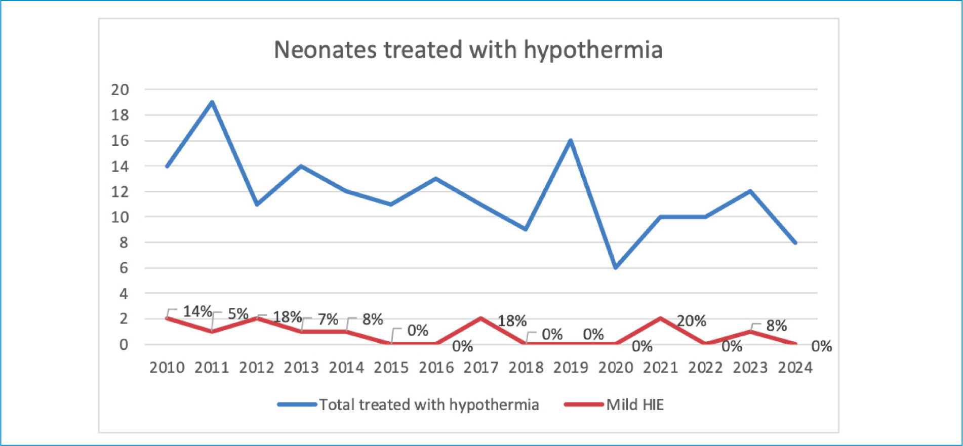
Figure 1. Annual distribution of neonates with HIE treated with therapeutic hypothermia.
All mild HIE neonates were outborn: nine were born in the Lisbon region, two in the Algarve, and one in the Azores islands.
Demographic and clinical characteristics
Most of the mothers were healthy and no complications during pregnancy were observed. All pregnancies were singleton. All neonates were born over 36 weeks of gestational age, and no significant prenatal problems were detected. A perinatal sentinel event was identified in seven cases. Additional birth-related variables are described in table 2.
Table 2. Pregnancy and delivery characteristics of neonates with mild HIE treated with therapeutic hypothermia
| Variable | Case | |||||||||||
|---|---|---|---|---|---|---|---|---|---|---|---|---|
| 1 | 2 | 3 | 4 | 5 | 6 | 7 | 8 | 9 | 10 | 11 | 12 | |
| Gender | M | M | F | F | F | M | F | M | M | M | F | F |
| BW (g) | 3530 | 3535 | 3450 | 2436 | 2070 | 3680 | 2046 | 3170 | 4070 | 3100 | 3145 | 2460 |
| GA (w) | 38 | 40 | 41 | 39 | 37 | 40 | 39 | 37 | 38 | 40 | 40 | 37 |
| Mode of delivery | Vacuum | Vacuum | Forceps | Eutocic | C-section | Eutocic | Vaginal breech | C-section | C-section | C-section | Vacuum | C-section |
| CTG | Normal | – | Suspicious | Normal | – | Suspicious | – | – | Suspicious | Suspicious | Normal | – |
| Amniotic fluid | Clear | Meconium | Meconium | Clear | – | Clear | Clear | Bloody | Meconium | Meconium | Clear | – |
| Sentinel event | Shoulder dystocia | None | Nuchal cord | Placental abruption | Placental abruption | Nuchal cord | None | Placental abruption | None | None | None | Placental abruption |
| DR resuscitation | I | I | I + CM + RD | I + CM + RD | I + CM + RD | I + CM + RD | I | I | I | I + CM + RD | I | I |
| Apgar score | 5-5-7 | 1-4-8 | 2-5-7 | 0-4-7 | 1-4-5 | 1-2-4 | – | 2-6-8 | 2-4-7 | 1-2-5 | 1-3-4 | 3-6-8 |
| pH* | 7.00 | 7.08 | 6.75 | 7.05 | – | 6.90 | 6.74 | 6.8 | 6.8 | 6.97 | 7.07 | 6.8 |
| Base deficit (mmol/L)* | 20 | 14 | 22 | 20 | – | 24 | 27 | 24.6 | 19.8 | 15 | 23.6 | Unmeasurable |
*Worst pH and base deficit in the first hour of life.
BW: birth weight; CM: cardiac massage; CTG: cardiotocography; DR: delivery room; F: female; g: grams; GA: gestational age; M: male; RD: resuscitation drugs; w: weeks.
All neonates required resuscitation at birth. Epinephrine was administered during resuscitation in five cases. One neonate underwent prolonged resuscitation (> 10 minutes). The median Apgar scores at one, five, and ten minutes were one, four, and seven, respectively. The mean pH in the first hour of life was 6.91 (range: 6.74-7.08). The base deficit ranged from unmeasurable (one case) to 27 mmol/L. Additional data relating to respiratory and cardiovascular support, complications, and medications administered during and after the TH protocol is presented in table 3. We point out that the protocol for empiric coverage for early-onset neonatal sepsis was changed during the studied period (the ampicillin plus cefotaxime regimen was changed to ampicillin plus gentamicin).
Table 3. Respiratory and cardiovascular support, complications, and medications administered in neonates with mild HIE during therapeutic hypothermia
| Variable | Case | |||||||||||
|---|---|---|---|---|---|---|---|---|---|---|---|---|
| 1 | 2 | 3 | 4 | 5 | 6 | 7 | 8 | 9 | 10 | 11 | 12 | |
| Respiratory support* | NIV only | IV | IV | IV | IV | IV | IV | NIV only | IV | IV | IV | NIV only |
| Antibiotics | Other regimen | AMP + CEF | AMP + CEF | AMP + CEF | AMP + CEF | AMP + CEF | AMP + CEF | AMP + GEN | AMP + CEF | AMP + GEN | AMP + GEN | AMP + GEN |
| Cardiovascular support | Inotrope(s) | Inotrope(s) | Inotrope(s) | None | Inotrope(s) | None | Inotrope(s) | Fluid bolus | Inotrope(s) | None | Inotrope(s) | Inotrope(s) |
| Acute kidney injury | Yes | No | No | No | No | No | No | No | No | No | No | No |
| Hypoglycemia | No | No | No | No | No | No | No | No | No | No | No | No |
| Coagulopathy | Yes | No | No | No | Yes | No | Yes | No | Yes | No | No | No |
| Length of stay (days) | 24 | 20 | 11 | 14 | 17 | 12 | 19 | 13 | 22 | – | 11 | 11 |
*IV occurred any time during hospitalization, prior and/or subsequent to NIV.
AMP: ampicillin; CEF: cefotaxime; GEN: gentamicin; IV: invasive ventilation; NIV: non-invasive ventilation.
Cooling characteristics
Most neonates were started on passive hypothermia within the first hour of life (median: one hour; range: 1-5 hours), and all were started on active hypothermia within the first 12 hours of life (median: 7.5 hours; range: 3-11 hours).
Neurological findings and neuromonitoring
In our center, the Thompson score is applied on admission and daily during TH to describe encephalopathy. The Sarnat classification was retrospectively estimated from available records. The median Thompson scores at admission, 24 hours, 48 hours, and at the end of rewarming were five, six, seven, and eight, respectively. All neonates were treated with morphine, and in two patients, midazolam or dexmedetomidine were also administered (Table 4). The morphine infusion dose had to be titrated to 15 mcg/kg/hour in two patients and up to 20 mcg/kg/hour in seven patients.
Table 4. Neuromonitoring, neurological findings during therapeutic hypothermia, and short-term neurological outcomes in neonates with mild HIE
| Variable | Case | |||||||||||
|---|---|---|---|---|---|---|---|---|---|---|---|---|
| 1 | 2 | 3 | 4 | 5 | 6 | 7 | 8 | 9 | 10 | 11 | 12 | |
| TS at admission | 0 | 0 | 5 | 5 | 6 | 6 | 4 | 3 | 5 | 5 | 6 | 3 |
| Sedation/analgesia (ug/kg/h) | MOR (< = 10) | MOR (> 10) | MOR (> 10) | MOR (> 10) | MOR (> 10) | MOR (> 10) | MOR (< = 10) | MOR (> 10) + MDZ | MOR (> 10) | MOR (> 10) | MOR (> 10) + DEX | MOR (< = 10) |
| Anticonvulsants | None | PHE | PHE | None | PHE | PHE | PHE | PHE | None | None | PHE + MDZ | None |
| aEEG* | Normal | Normal | Normal | Normal | Moderately abnormal | Moderately abnormal | Moderately abnormal | Suppression | – | Normal | Moderately abnormal | Normal |
| Electrical seizure on aEEG | No | Yes | Yes | No | Yes | No | No | No | No | No | Yes | No |
| Exclusive oral feeding at day 14 | Yes | Yes | Yes | Yes | No | Yes | No | Yes | No | Yes | Yes | Yes |
| Auditory evoked potentials | Normal | – | Normal | Normal | Normal | Normal | Normal | Normal | – | Normal | Normal | Normal |
| EEG at discharge | Normal | – | Abnormal | Normal | – | Normal | Normal | Abnormal | – | Normal | Abnormal | – |
| Clinical seizures at discharge | No | No | No | No | No | No | No | No | No | No | No | No |
*Worst aEEG during hospitalization.
DEX: dexmedetomidine; MDZ: midazolam; MOR: morphine; PHE: phenobarbital; TS: Thompson score.
Three infants had suspected clinical seizures at the referring hospital. In one infant, TH was initially considered not indicated; however, seizures occurred 12.5 hours after birth during rewarming, leading to the reinstatement of TH.
Electrical seizures were documented in three additional infants during treatment. In total, seven infants received anticonvulsants, with phenobarbital administered in every case, and a combination of phenobarbital and midazolam in one case.
On admission, the aEEG was normal in most neonates, while four had a moderately abnormal background. In each of the patients with an altered aEEG, the background normalized during TH (in two at 48 hours, one at 60 hours, one at 14 hours, and one at 16 hours). In one case, the aEEG pattern worsened during treatment, progressing to a burst-suppression pattern before eventually normalizing (Table 4).
Cranial ultrasound
The cranial ultrasound showed evident or equivocal grey-white matter differentiation in four neonates, a resistive index of < 0.55 in three, and no signs of cerebral edema in any of the cases.
MRI
Brain MRI was performed at a median age of 11.5 days (range: 7-16). Six neonates showed normal findings or only a small subdural hemorrhage (Weeke score 0-1). One patient showed signs of a left-sided ischemic infarct. Another patient exhibited cortical highlighting (Weeke score 1), and two showed equivocal myelination of the posterior limb of the internal capsule (PLIC) (Weeke score 1-2). One neonate showed basal ganglia lesions, Rolandic cortical highlighting, and equivocal PLIC (score 5). No MRI results were available for one patient.
Evaluation at discharge
The median NICU stay was 14 days (range: 11-24). Three neonates were transferred back to their original NICU and the remainder were discharged home. Neurological examination at discharge was normal in all but one infant with hypotonia and hyporeflexia. No seizures or anticonvulsant therapy were reported at discharge. Most neonates had a normal EEG and auditory evoked potentials. By day 14, only three required tube feeding, but all were discharged on oral feeding (Table 4). No deaths occurred. One neonate was diagnosed with ischemic cerebral infarct on MRI, and no other alternative diagnoses were identified.
Discussion
In this study, we describe our 14-year experience with therapeutic hypothermia in neonates with mild HIE in a level III NICU in Portugal. Although the overall profile of treated infants remained relatively stable, we observed a trend toward including neonates with mild symptoms or requiring only respiratory resuscitation. This pattern is consistent with international reports, reflecting both increasing awareness of the potential risks of mild HIE and growing confidence in the safety of TH in routine practice. Nevertheless, the proportion of mild HIE infants who underwent TH in our cohort was substantially lower than the 22% reported in a meta-analysis from seven high-income countries.2,10
A sentinel event was identified in 58% of our cases, higher than the one-third typically reported.15 This may have influenced the decision to include some infants with mild symptoms in the TH protocol. Cardiovascular and respiratory support requirements were notable, with invasive ventilation needed in 75% and inotropic support in 67% of cases. Non-invasive ventilation was successfully used in several infants, in line with the current trend toward less invasive strategies to reduce complications.
Sedation was also a relevant challenge: nine out of 12 infants required high-dose morphine or a second agent, likely reflecting milder encephalopathy and greater discomfort associated with cooling. The need for higher opioid doses raises concerns about toxicity and highlights the potential risks of TH in mild HIE, including maternal separation, breastfeeding interference, and increased susceptibility to nosocomial infection. These factors may partly explain why current studies have not demonstrated a clear benefit.
On admission, four infants had a moderately abnormal aEEG, which likely influenced the decision to initiate TH, as our initial local guideline incorporated aEEG as a treatment criterion following the TOBY protocol.16 aEEG has also been used in clinical trials and in practice to help select infants with mild HIE for cooling.17–19 In this context, discontinuity, asymmetry, or a poor sleep-wake cycle may suggest an increased risk of an adverse outcome, although a normal aEEG does not exclude this.7,20,21 In our cohort, background activity normalized within 48 hours in all but one infant, generally indicating a favorable prognosis, though less reassuring features were observed in four cases. Four infants had electrographic seizures; in one, the aEEG pattern deteriorated to burst-suppression before recovering, corresponding to the most severe MRI injury. Among the seizure group, one infant had an ischemic infarct, another had mild cortical highlighting, and two had normal MRI findings.
MRI abnormalities were present in 41,7% of infants, mostly mild, but one neonate showed a significant basal ganglia lesion (Weeke score 5). MRI is a reliable predictor of long-term neurodevelopment, and studies consistently report abnormalities in 34-66% of mild HIE cases, a rate comparable to moderate or severe HIE.1,2,7,20–22 In mild HIE, injury patterns are usually less severe (periventricular edema, punctate white matter lesions, and minimal cortical highlighting) and may not translate into significant long-term poor outcomes, but misclassifying these subtle changes as abnormal can overestimate the prognosis of adverse outcomes. In fact, significant basal ganglia or thalamic injury on T1- and T2-weighted images is rare, occurring in < 4% of cases.6,23
The classification of HIE severity within the first six hours remains a major challenge. Neurological signs fluctuate over time, inter-observer variability is significant, and there is no universally accepted definition of perinatal hypoxia-ischemia or of the grading of encephalopathy. Reported progression rates from mild to more severe encephalopathy varies widely across studies (1.6% in PRIME, 12% in Gagne-Loranger et al., 26.7% in MARBLE, and up to 50% in ICE).5,19,24 This uncertainty may explain why some infants with mild HIE and additional risk factors, such as an abnormal aEEG, seizures, or sentinel events, are cooled in clinical practice. In our cohort, most cooled infants with mild HIE had one of these features and international and national guidelines appear to be generally followed.
Ultimately, deciding which infants with mild HIE should receive TH is complex. Two ongoing randomized trials may soon provide crucial evidence. The COOL PRIME study (NCT04621279) is evaluating TH versus normothermia with neurodevelopmental outcomes at two years, while the COMET trial (NCT03409770) is testing normothermia versus TH for 48 or 72 hours, using magnetic resonance spectroscopy at two weeks as a surrogate outcome. These results will be critical to clarify the role of TH in this large group of infants.
Our study has limitations. Its retrospective design introduces variability in assessment and documentation, and the absence of a control group of untreated mild HIE infants prevents conclusions regarding efficacy or safety. Despite these limitations, our findings reflect real-world practice and highlight the importance of aligning clinical decisions with current evidence and guidelines, particularly given the risks of therapy.
In summary, the management of mild HIE remains an area of uncertainty. Early classification within the first six hours is complex and often unreliable, as some infants initially presenting with mild signs later evolve to more severe stages. Although increasing evidence links mild HIE with brain injury and adverse neurodevelopmental outcomes, current data does not justify routine use of TH in this group, which represents almost half of all HIE cases. The balance between potential benefit and possible harm is still unclear, highlighting the urgent need for well-designed clinical trials. Improving early risk stratification through refined neurological assessment, neurophysiological monitoring, and emerging biomarkers will be key. As the use of TH in mild HIE expands, studies such as ours add to the understanding of real-world practice and outcomes, and reinforce the importance of cautious, evidence-based decision-making.
Author contributions
J. Fortuna: design, data curation, formal analysis, methodology, software, visualization, writing of the original draft, review and editing; F. Proença: design, formal analysis, methodology, validation, review and editing; A. Graça: design, data curation, formal analysis, investigation, methodology, supervision, validation, visualization, review and editing; I. Sampaio: design, formal analysis, methodology, validation, review and editing.
Previous presentations
52º Congresso Português Neonatologia, Eurostars Oasis Plaza, Figueira da Foz. November 29, 2024.
Funding
None.
Conflicts of interest
None.
Ethical considerations
Protection of humans and animals. The authors declare that no experiments involving humans or animals were conducted for this research.
Confidentiality, informed consent, and ethical approval. The authors have obtained approval from the Ethics Committee for the analysis of routinely obtained and anonymized clinical data, so informed consent was not necessary. Relevant guidelines were followed.
Declaration on the use of artificial intelligence. The authors declare that no generative artificial intelligence was used in the drafting of this manuscript.
