

Keypoints
What is known
– In KBGS, the value of a dysmorphological examination should not be undervalued. Targeted analysis of the ANKRD11 gene confirms most clinical suspicions, but WES is the preferred option for differential diagnosis. In most cases, developmental delay/intellectual disability are mild, but behavioral changes seem to be quite prevalent and should be valued and addressed.
What is added
– This study documents a total of 15 previously unreported pathogenic or likely pathogenic sequence variants in the ANKRD11 gene and characterizes 35 individuals with a confirmed molecular diagnosis of KBGS, including a family with three affected generations.
Introduction
KBG syndrome (MIM#148050; KBGS) is an autosomal dominant syndromic developmental delay/intellectual disability disorder, caused by haploinsufficiency of the ANKRD11 (ankyrin repeat domain-containing protein 11) gene or deletions of the 16q24.3 region encompassing ANKRD11. Most cases are de novo.
ANKRD11 was reported as the causative gene for KBGS in 2011.1 It encodes a 2663 amino acid-long protein containing five ankyrin repeats, two repression domains (repression domain 1, RD1; repression domain 2, RD2) and one activation domain (AD), which plays a key role in brain development.2 The ANKRD11 protein is a chromatin regulator that has previously been shown to control neural stem cell fates via modulation of histone acetylation.3 Recent studies suggest that ANKRD11 gene variants disrupting RD1 and RD2, or RD2 alone, are likely to cause more severe developmental delay/intellectual disability.4
There is no consensus on the diagnostic clinical criteria for KBGS, although several have been suggested.5,6,7 In addition to developmental delay/intellectual disability, the main manifestations are macrodontia (especially of the permanent upper central incisors), typical facial appearance and post-natal short stature (length and/or height < 3rd centile). The facial gestalt is characterized by a triangular face, synophrys, widely spaced eyes, broad eyebrows, prominent ears, a bulbous nose and anteverted nares. Additional findings include chronic or recurrent otitis media, hearing loss (mostly conductive, but also mixed and sensorineural), palatal anomalies, hair anomalies, brachydactyly, a large anterior fontanelle with delayed closure, costovertebral anomalies, scoliosis, delayed bone age, electroencephalogram abnormalities with or without seizures, feeding difficulties and cryptorchidism in males. There is significant variability in clinical findings among and within families, and KBGS is likely an underdiagnosed cause of developmental delay/intellectual disability, since many features are nonspecific, especially before eruption of the permanent dentition.1
Some large cohorts of patients with developmental delay/intellectual disability studied by whole exome sequencing (WES) identified patients with ANKRD11 variants and a non-classic Cornelia de Lange syndrome (CdLS) phenotype, indicating a clinical overlap between KBGS and CdLS. This is not surprising since ANKRD11, like proteins linked to CdL, is involved in chromatin regulation, controlling gene expression during neural development.8,9,10
In this paper we describe the clinical and molecular characterization of 35 Portuguese KBGS patients identified in four national medical genetics centers. We compare our data with other recently published cohorts.
Methods
This is a retrospective review of clinical and molecular data from Portuguese KBGS patients. We obtained information on a total of 35 patients, including 17 females and 18 males currently between three and 73 years of age (average age of 18.9 years old). There were three families with more than one affected individual (F1-F3), representing nine patients in total. In one family there were three affected generations (F3). A total of 16 patients were diagnosed in Unidade Local de Saúde Santa Maria, 13 in Unidade Local de Saúde de Coimbra, four in Unidade Local de Saúde de Santo António and two in Unidade Local de Saúde de São José. This study was approved by the Ethics Committee of the Lisbon Academic Medical Center.
Data was collected through a form filled in by the clinical geneticists of each center. Informed consent for anonymous publication of molecular and clinical information was obtained in every case. The photographs shared in this article were specifically consented to. Molecular data was reviewed in Alamut® Software, a bioinformatics application for genetic analysis. Only patients with a confirmed molecular diagnosis were included (patients with variants of uncertain significance in the ANKRD11 gene were excluded). Patients’ photos were analyzed in Face2Gene® Software, a computer-aided phenotyping tool.
Results
Molecular results
Of the 35 patients included in this cohort, 32 (P1-32) had an ANKRD11 sequence variant and three (P33-35) had a large deletion involving the ANKRD11 gene.
A total of 23 different sequence variants were identified (Table 1 and Fig. 1), of which 15 were previously unreported in the HGMD® (Human Gene Mutation Database). Based on ACMG (American College of Medical Genetics and Genomics) variant classification criteria, we uncovered 23 variants classified as pathogenic or likely pathogenic, including 20 frameshifts, two missense and one splice site variants. Segregation studies in familial cases concluded the variant had a maternal origin in two families and was of paternal origin in one case.
Table 1. ANKRD11 sequence variants found in the present series of KBGS patients
| Family | Patient | Variant coordinates NM_013275.6(ANKRD11) |
Exon | Protein change | Nature of mutation | Classes | HGMD | Origin |
|---|---|---|---|---|---|---|---|---|
| P1 | c.226+1G>T | Int | p? | Splicing | LP | NR | Unknown | |
| P2 | c.866dup | 8 | p.Tyr289* | Frameshift | LP | NR | De novo | |
| P3 | c.1617dup | 9 | p.Asp540Argfs*39 | Frameshift | LP | NR | De novo | |
| P4 | c.1670del | 9 | p.Pro557Argfs*17 | Frameshift | LP | NR | De novo | |
| F1 | P5 | c.1902_1905del | 9 | p.Lys635Thrfs*17 | Frameshift | P | NR | Paternal |
| P6 | Unknown | |||||||
| P7 | c.1903_1907del | 9 | p.Lys635Glnfs*26 | Frameshift | P | R | De novo | |
| P8 | c.1903_1907del | 9 | p.Lys635Glnfs*26 | Frameshift | P | R | Unknown | |
| P9 | c.1903_1907del | 9 | p.Lys635Glnfs*26 | Frameshift | P | R | De novo | |
| P10 | c.1920_1921insT | 9 | p.Lys641* | Frameshift | LP | NR | De novo | |
| P11 | c.2175_2178del | 9 | p.Asn725Lysfs*23 | Frameshift | P | NR | De novo | |
| P12 | c.2329_2332del | 9 | p.Glu777Argfs*5 | Frameshift | P | NR | De novo | |
| P13 | c.2344_2345dup | 9 | p.Leu782Phefs*2 | Frameshift | LP | NR | De novo | |
| P14 | c.2398_2401del | 9 | p.Glu800Asnfs*62 | Frameshift | P | R | De novo | |
| P15 | c.2512C>T | 9 | p.Arg838* | Frameshift | P | R | De novo | |
| P16 | c.3201dup | 9 | p.Asp1068Argfs*34 | Frameshift | LP | NR | De novo | |
| P17 | c.3309del | 9 | p.Asp1104Metsf*214 | Frameshift | P | R | De novo | |
| P18 | c.3309dup | 9 | p.Asp1104Argfs*2 | Frameshift | LP | R | Unknown | |
| P19 | c.3345_3346insTT | 9 | p.Ala1116Leufs*203 | Frameshift | LP | R | De novo | |
| P20 | c.3770_3771del | 9 | p.Lys1257Argfs*25 | Frameshift | P | NR | De novo | |
| P21 | c.3770_3771del | 9 | p.Lys1257Argfs*25 | Frameshift | P | NR | Unknown | |
| F2 | P22 | c.3786_3789del | 9 | p.Lys1262Asnfs*55 | Frameshift | P | NR | Maternal |
| P23 | Unknown | |||||||
| F3 | P24 | c.4384dup | 9 | p.Arg1462Lysfs*92 | Frameshift | P | R | Maternal |
| P25 | Unknown | |||||||
| P26 | Maternal | |||||||
| P27 | Maternal | |||||||
| P28 | Maternal | |||||||
| P29 | c.6841C>T | 9 | p.Gln2281* | Frameshift | LP | NR | De novo | |
| P30 | c.7238_7249delinsGT | 9 | p.Gln2413Args*75 | Frameshift | LP | NR | De novo | |
| P31 | c.7535G>A | 10 | p.Arg2512Gln | Missense | LP | R | Unknown | |
| P32 | c.7814T>G | 13 | p.Leu2605Arg | Missense | LP | NR | De novo |
F1-3: family 1–3; int – intronic; LP: likely pathogenic; P: pathogenic; P1–32 – patient 1–32; HGMD: human gene mutation database; NR: not reported; R: reported.
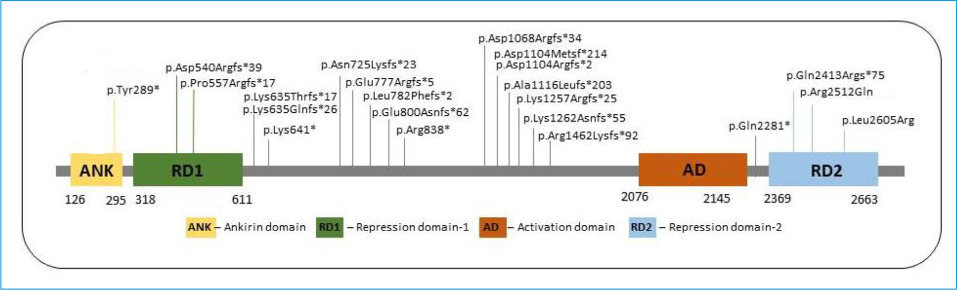
Figure 1. Distribution of the ANKRD11 variants at the protein level: a schematic representation of the ANKRD11 protein with the representation of two repression domains (repression domain 1, RD1; repression domain 2, RD2) and one activation domain (AD).
In most patients, molecular analysis was performed by targeted testing of the ANKRD11 gene (46.5%; [14/30]), but some were studied by whole exome sequencing (27%; [8/30]) or using a multigene NGS panel including ANKRD11 (6.5%; [2/30]). Familial cases were tested for the familial variant (20%; [6/30]). ANKRD11 deletions were identified by an array CGH (Comparative Genomic Hybridization) test.
Clinical results
We report the clinical manifestations specifically asked about in the form filled in by clinical geneticists from each center. The results are presented as percentages corresponding to the proportion of positive individuals in the total number of responses obtained for each question/manifestation. Individual data pertaining to the main clinical features in table 3 are shown in Supplementary Table 1 and to dysmorphisms in table 4 in Supplementary Table 2.
PRENATAL, BIRTH AND NEONATAL COMPLICATIONS
Intrauterine growth restriction (IUGR) was detected in 17% (4/24) of the patients, and 14% (4/28) presented other nonspecific prenatal abnormalities detected by fetal ultrasound, such as single umbilical artery, mild renal pelvis dilatation and polyhydramnios. All but three patients were born at term. In the neonatal period, 37% (11/30) had feeding difficulties, 27% (7/26) had hypotonia and 16% (5/32) had respiratory distress.
GROWTH PARAMETERS
Short stature (below 3rd centile or –2SD) was observed in 40% (14/35) of the patients. Additionally, 9% (3/35) were between the 3rd and 15th centile, equating to almost half of the patients with a stature below the 15th centile. Microcephaly (below the third centile or –2SD) was present in 7% (2/28).
NEURODEVELOPMENT AND THE CENTRAL NERVOUS SYSTEM (CNS)
All of the patients had global developmental delay (31/31) and learning difficulties (33/33). Out of the 12 patients who had a formal evaluation, 11 (92%) had an IQ (intelligence quotient) below 80: 44% (4/9) had borderline intellectual disability (ID) (IQ 70-79), 44% (4/9) had mild ID (IQ 50-69), 11% (1/9) had moderate ID (IQ 36-49) and one patient had an IQ of 82. We also evaluated the prevalence of two behavioral problems: ADHD (attention deficit hyperactivity disorder) was present in 67% (14/21) of the patients and autism in 10% (2/20).
Table 2. Genome coordinates of ANKRD11 deletions found in the present series of KBGS patients
| Patient | Genome coordinates (hg19) of ANKRD1 deletions | Classes | Origin |
|---|---|---|---|
| P33 | Chr16: 88885164_89520963 | p | De novo |
| P34 | Chr16: 88926780_89434598 | p | De novo |
| P35 | Chr16: 89394003_89561269 | p | De novo |
P33–35: patient 33–35; P: pathogenic.
CNS abnormalities were present in 43% (6/14) of the patients who had brain imaging, including one patient with an enlarged cisterna magna, two patients with dysgenesis of the corpus callosum and three patients with other nonspecific abnormalities. Seizures were reported in 19% (5/26) of the patients.
VISION AND HEARING
A total of 24% (6/25) of the patients had strabismus, 26% (6/23) had astigmatism, 21% (5/24) had myopia and 4% (1/23) had hypermetropia.
Half of the patients (14/28) had hearing loss, mostly classified as conductive (57%; 8/14). In this line, 57% (13/23) of the patients had a personal history of recurrent otitis media and 50% (10/20) had undergone ENT (ear, nose and throat) surgery. Only one patient had a cleft palate.
CARDIAC AND UROGENITAL
Cardiac anomalies included ventricular septal defects, reported in 17% (4/24) of the patients, auricular septal defects in 12% (3/25) and a patent foramen ovale in 9% (2/22). Cryptorchidism was present in 33% (4/12).
SKELETAL
A large anterior fontanelle with delayed closure was reported in 12% (2/17) of the patients. Costovertebral anomalies were identified in the chest X-ray of three patients (14%; 3/22), one consisting of cervical ribs and posterior fusion defects of S1 (Fig. 2). Other skeletal anomalies included clinodactyly/brachydactyly in 85% (28/33), short toes in 40% (10/25), syndactyly of toes in 37% (7/19), a widely spaced gap between the first and second toes in 18% (4/22) and pes planus in 15% (3/20).
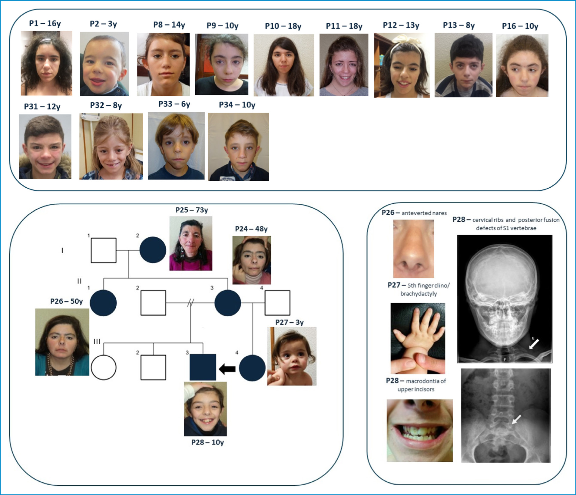
Figure 2. Facial features and other clinical signs of KBGS patients included in this series. A: individuals with pathogenic (P)/likely pathogenic (LP) sequence variants or deletions of the ANKRD11 gene. All indicated ages correspond to the last clinical evaluation; B: family tree of Family 3. The arrow indicates the index case who was then studied by targeted sequencing of the ANKRD11 gene. The sequence variant identified was then tested for in the remaining elements of the family, allowing the diagnosis of KBGS in his maternal half-sister, mother, aunt and grandmother; C: some specific clinical signs of KBG syndrome found in elements of Family 3.
CRANIOFACIAL
Photographs of patients belonging to this cohort are presented in figure 2. A typical craniofacial gestalt was recognized in 76% (26/34) of the patients. The phenotype recognition bioinformatics tool Face2Gene® was applied in 16 patients, of whom 14 had a high score for KBGS, one a moderate score and one a low score. In 15% (4/26), Cornelia de Lange syndrome was first suspected.
The most frequently reported dysmorphisms were prominent macrodontia of the upper incisors in 81% (21/26), a prominent nasal bridge in 79% (26/33), a broad nasal tip in 77% (27/35), synophrys in 62% (21/34), a wide triangular face in 58% (19/33), long palpebral fissures in 48% (14/29), anteverted nares in 50% (17/34), a long philtrum in 40% (12/30) and large ears in 38% (10/26).
Discussion
Discussion of the molecular results
We highlight that almost half (46.5%) of the molecular diagnoses were made by targeted testing of the ANKRD11 gene. This high percentage is explained, on the one hand, by KBGS having a recognizable gestalt, and, on the other hand, because most patients were diagnosed prior to the widespread clinical use of multigene panels and whole exome sequencing (WES). In the meantime, WES has been gradually adopted in the investigation of (syndromic) ID. This option is very cost-effective given the wide phenotypic overlap and broad genetic heterogeneity of ID, so it is to be expected that in the future the large majority of KBGS diagnoses will be made in extended studies.
Discussion of the clinical results
The main purpose of this study was to perform the clinical characterization of Portuguese KBGS patients and compare it with the literature, highlighting any features that may have been previously undervalued or unreported. We have collected clinical data from 35 individuals with a molecular diagnosis of KBGS. In table 3, this data is compared with patients from four cohorts published in recent years.11,12,13,14,15 Four of these cohorts include between 12 and 31 patients, which is fewer than in our series, reinforcing the relevance of our contribution to the clinical characterization of this condition. The most recent study includes 67 patients, representing the largest published cohort to date.15
Table 3. Comparison of the prevalence of clinical features between our cohort and four recent series of KBGS patients11–15
| Manifestation | Our cohort | Parenti I. et al. | Kutkowska-Kazmierczak et al. | Gnazzo M. et al. | Scarano E. et al. | Martínez-Cayuelas, et al. |
|---|---|---|---|---|---|---|
| Pregnancy complications | ||||||
| IUGR | 17% (4/24) | Not rated | Not rated | Not rated | 8% (1/12) | 22% (15/58) |
| Prenatal abnormalities | 14% (4/28) | Not rated | Not rated | Not rated | 16% (2/12) | Not rated |
| Neonatal complications | ||||||
| Feeding difficulties | 37% (11/30) | Not specific to the neonatal period: 30% (7/23) | Not specific to the neonatal period: 60% (14/23) | Not specific to the neonatal period: 36% (11/31) | 8% (1/12) | Rated as pregnancy and perinatal complications: 39% (18/46) |
| Hypotonia | 27% (7/26) | Not rated | Not rated | Not rated | Not rated | |
| Respiratory distress | 16% (5/32) | Not rated | Not rated | Not rated | Not rated | |
| Growth and head | ||||||
| Short stature (< P3) | 40% (14/35) | 26% (6/23) (< –2SD) | 35% (8/23) (< –2SD) | 58% (18/31) | 75% (9/12) (< –1SD) | 52% (28/54) (< P10) |
| Microcephaly (< P3) | 7% (2/28) | 13% (3/23) (< –3SD) | Not rated | Not rated | 25% (3/12) | 21% (10/48) |
| Neurodevelopment | ||||||
| GDD | 100% (31/31) | 100% (22/22) | DD/ID: 74% (17/23) Speech delay: 85% (18/21) |
Delayed motor milestones: 52% (16/31) Speech delay: 77% (23/30) |
83% (10/12) | Motor delay: 58% (32/55) Speech delay: 72% (36/50) |
| ID (IQ < 80) | 92% (11/12) | 85% (17/20) | Not rated | 83% (10/12) | 83% (46/56) | |
| Learning difficulties | 100% (33/33) | Not rated | Not rated | 87% (26/30) | Not rated | Not rated |
| ADHD | 67% (14/21) | Behavioral problems: 68% (15/22) |
86% | Not rated | Not rated | 63% (31/49) |
| Autism | 10% (2/20) | Not rated | Not rated | Not rated | 41% (22/53) | |
| Central nervous system | ||||||
| Reported abnormalities | 43% (6/14) | 22% (4/18) | Not rated | 7% (1/15) | 80% (8/10) | Abnormal MRI: 38.3% (18/47) |
| Epilepsy | 19% (5/26) | 26% (6/23) | Not rated | 16% (5/31) | 42% (3/7) | 25% (15/61) |
| Eyes | ||||||
| Strabismus | 24% (6/25) | Not rated | Not rated | 13% (4/13) | Not rated | 18% (9/49) |
| Myopia | 21% (5/24) | Not rated | Not rated | Not rated | Not rated | Visual problems: 43% (23/54) |
| Hypermetropia | 4% (1/23) | Not rated | Not rated | Not rated | Not rated | |
| Astigmatism | 26% (6/23) | Not rated | Not rated | Not rated | Not rated | |
| Ear nose throat | ||||||
| Recurrent otitis media | 57% (13/23) | Hearing problems: 31% (7/22) |
Not rated | Not rated | Not rated | 42% (22/53) |
| Hearing loss | 50% (14/28) Conductive: 57% (8/14) |
Not rated | 19% (6/31) Sensorineural: 7% (2/31) Conductive: 16% (5/31) |
25% (3/12) | 44% (25/57) Sensorineural: 35% (6/17) Conductive: 53% (9/17) Mixed: 12% (2/17) |
|
| ENT surgery | 50% (10/20) | Not rated | Not rated | Not rated | Not rated | Not rated |
| Cleft palate | 5% (1/21) | Not rated | Not rated | Not rated | Not rated | Not rated |
| Cardiac | ||||||
| Auricular septal defect | 12% (3/25) | Cardiac anomalies: 17% (4/23) |
Not rated | Not rated | Heart problems: 41% (5/12) |
Congenital heart defects: 28% (16/58) |
| Ventricular septal defect | 17% (4/24) | Not rated | Not rated | |||
| Patent foramen ovale | 9% (2/22) | Not rated | Not rated | |||
| Urogenital | ||||||
| Cryptorchidism | 33% (4/12) | 25% (3/12) | Not rated | 13% (2/15) | 25% (2/8) | 36% (12/33) |
| Skeletal | ||||||
| Large/delayed closure of anterior fontanel | 12% (2/17) | Not rated | 56% (13/23) | Not rated | 8% (1/12) | Not rated |
| Costovertebral anomalies | 14% (3/22) | Not rated | Not rated | Not rated | 91% (11/12) | Not rated |
| 5th finger clino/brachydactyly | 85% (28/33) | 70% (16/23) | Not rated | 23% (7/31) | Hand clinodactyly 100% (12/12) Hand Brachydactyly 83% (10/12) |
57% (27/47) |
| Feet brachydactyly | 40% (10/25) | Brachydactyly: 47% (11/23) |
Not rated | Hand brachydactyly: 68% (21/31) | Not rated | Not rated |
| Toes syndactyly | 37% (7/19) | 23% (5/22) | Not rated | 2nd-3rd toe syndactyly: 10% (3/31) | Not rated | Not rated |
| Widely spaced gaps between 1st and 2nd toes | 18% (4/22) | Not rated | Not rated | Not rated | Not rated | Not rated |
| Pes planus | 15% (3/20) | Not rated | Not rated | Not rated | Not rated | Not rated |
ADHD: attention deficit hyperactivity disorder; ENT: ear, nose and throat; GDD: global developmental delay; ID: intellectual disability; IUGR: intrauterine growth restriction.
Table 4. Comparison of the prevalence of dysmorphisms between our cohort and four recent series of KBGS patients11–15
| Dysmorphisms | Our cohort | Parenti I. et al. | Kutkowska-Kazmierczak et al. | Gnazzo M. et al. | Scarano E. et al. | Martínez-Cayuelas et al. |
|---|---|---|---|---|---|---|
| Typical craniofacial gestalt of KBG | 76% (26/34) | 80% (17/21) | Not rated | Not rated | Not rated | Not rated |
| First suspected as Cornelia de Lange | 15% (4/26) | 34% (8/23) | Not rated | Not rated | Not rated | Not rated |
| Wide triangular face | 58% (19/33) | 60% (14/23) | Not rated | Not rated | Coarse face: 100% (12/12) | 71% (37/52) |
| Long palpebral fissures | 48% (14/29) | Not rated | Not rated | Not rated | Not rated | Not rated |
| Synophrys | 62% (21/34) | 65% (15/23) | Not rated | 90% (28/31) | Not rated | 38% (23/60) |
| Large ears | 38% (10/26) | Not rated | Not rated | 23% (7/31) | 100% (12/12) | 56% (28/50) |
| Prominent nasal bridge | 79% (26/33) | Not rated | Not rated | Not rated | Nose abnormalities: 100% (12/12) |
38% (17/45) |
| Broad nasal tip | 77% (27/35) | 70% (16/23) | Not rated | 65% (20/31) | 59% (30/51) | |
| Anteverted nares | 50% (17/34) | 78% (18/23) | Not rated | Not rated | 57% (24/42) | |
| Long philtrum | 40% (12/30) | 83% (19/23) | Not rated | 90% (28/31) | 100% (12/12) | 57% (28/49) |
| Macrodontia of upper incisors | 81% (21/26) | 65% (15/23) | 52% (11/21) | 77% (23/30) | 100% (9/9) | 77% (35/47) |
The clinical features of Portuguese KBGS patients are overall in accordance with what has been previously described in the literature. Some of the manifestations evaluated in this cohort had not been previously valued or individually characterized in the other series. We found it pertinent to report these manifestations with the aim of contributing to greater clinical sensitivity in other patients.
Global developmental delay, learning difficulties and borderline-mild intellectual disability are among the main features of KBGS, consistently found in all series under comparison. We did not confirm the previously suggested hypothesis of more severe developmental delay/intellectual disability associated with variants disrupting RD1 and RD2, or RD2 alone.4 In fact, the only patient with moderate ID (P8) presents a variant located outside the functional domains of the gene. Behavioral issues, on the other hand, were not so well characterized in general, and would be worth exploring further. It is important to clarify whether these diagnoses reflect conclusions derived from formal clinical assessments or merely the identification of personality traits, as this distinction may significantly impact the reported prevalence of such manifestations. In our cohort, patients were evaluated in developmental clinics, supporting the assumption of a formal diagnosis of ADHD and/or autism. In contrast, Kutkowska-Kaźmierczak et al.12 reported a notably high prevalence of ADHD but referred primarily to psychomotor hyperactivity, which may explain the elevated rates observed in their study. Conversely, Martínez-Cayuelas et al.15 described a prevalence comparable to that observed in our cohort and included patients presenting either a formal diagnosis or phenotypic features consistent with these conditions. Nonetheless, the prevalence of ADHD appears to be considerable and underscores the need for the systematic and targeted evaluation of all individuals with KBGS. Autism was assessed only in the cohort described by Martínez-Cayuelas et al.,15 which included patients with a formal diagnosis of autism spectrum disorder and demonstrated a higher prevalence than that observed in our cohort.
Fewer than half of the patients in our cohort underwent brain imaging examinations, so it is likely that brain abnormalities are underdiagnosed. In fact, when compared with the literature, the prevalence is higher in the Scarano et al. cohort,14 most often with enlargement of the cisterna magna, which was also described in one of our patients. The most frequent brain anomaly in our patients was dysgenesis of the corpus callosum, which was present in two cases. These anomalies are unspecific, and brain MRI is not generally recommended, but should be considered, especially in patients with seizures and/or microcephaly.
Until recently, ocular manifestations in patients with KBGS had been scarcely investigated in the literature. To address this gap, Carter et al.16 conducted a study in 2023 specifically focused on these features and reported that, out of 40 KBGS patients, strabismus was observed in 22.5%, while astigmatism, myopia and hypermetropia were reported in 27.5%, 15% and 20% of patients, respectively. With the exception of hypermetropia, for which we observed a lower prevalence, the remaining findings are largely consistent with our results.
Similarly, hearing impairment in KBGS has been specifically evaluated in a recent study by Rhamati et al.,17 who reported that, out of 32 KBGS patients, a typical audiological profile was observed: predominantly bilateral conductive (81%), mild to moderate (84%) and stable (69%) hearing loss, with some audiological heterogeneity. Our findings are consistent with this characteristic profile of hearing loss. Importantly, Rhamati et al.17 also reported that more than half of the evaluated patients (55%) exhibited abnormalities on CT imaging, most frequently involving the ossicular chain. It should be noted that our cohort did not assess whether patients had undergone imaging studies, such as internal auditory canal MRI, which would have been highly relevant for drawing further conclusions. In view of these findings, regular hearing evaluation, including comprehensive audiological and radiological assessment, as well as ENT follow-up, is recommended for all patients with KBGS.
In this cohort, the particular type of cardiac malformation was evaluated, showing that the most frequent cardiac anomaly are septal defects (a ventricular septal defect in 17%, and an atrial septal defect in 12%). Therefore, if not previously performed, we also recommend a baseline cardiac evaluation at the time of diagnosis.
Identifying a large anterior fontanelle with delayed closure is dependent on the age at clinical observation and an attentive, directed examination. The Kutkowska-Kaźmierczak et al.12 series specifically evaluated less explored features of KBGS, including this characterization, thus it is not surprising that the prevalence in their cohort is higher than in our and the Scarano et al.14 series. Clinicians should be aware of this feature and actively look for it.
Likewise, we found it difficult to assess the prevalence of costovertebral abnormalities since it depends on the active search for these alterations, including skeletal radiography. In the Scarano et al.14 series, radiography was requested for all patients, so the prevalence of costovertebral abnormalities is much higher than in our series. However, the anomalies reported by Scarano et al.14 are identical to those identified in one of our patients: cervical ribs and posterior fusion defects of S1. The investigation of syndromic ID does not usually involve skeletal radiography. Also, short stature in KBGS is classically proportioned, which does not warrant skeletal radiography. The skeletal manifestations found in KBGS patients so far do not have functional implications or require any intervention. Therefore, we believe that radiography is not essential but contributes to the clinical characterization of these patients.
We individually assessed the prevalence of particular dysmorphisms in patients with KBGS. In general, it is consistent with the other series under comparison, especially that of Parenti et al.11 and Martínez-Cayuelas et al.,15 who endeavored to do the same. Macrodontia of the upper central incisors is the main common finding, but it pertains to the permanent dentition, so younger children may be underdiagnosed. The second most prevalent facial feature concerns the nose, which is described as having a prominent bridge, broad tip and anteverted nares. Both these features are usually present in patients with KBGS, but can be somewhat subjective, requiring a careful examination by a well-trained clinician.
It is also important to highlight that in our cohort, clinical geneticists believed most of the patients presented the typical gestalt of KBGS. Additionally, KBGS should be considered as a differential diagnosis of Cornelia de Lange syndrome, first suspected in 15% of the patients in our cohort.
The transition to adulthood may be particularly challenging, and most patients are expected to require ongoing support due to the impact of intellectual disability, even when mild, on achieving full independence. While some individuals may attain a degree of autonomy, the need for assistance remains the norm. Adult functioning in KBGS is further significantly affected by prevalent neurodevelopmental and psychiatric comorbidities, with only a minority achieving stable and remunerated employment.18,19 In this regard, it is worth mentioning that the adult individuals of F3 (the family with the highest number of patients) have achieved sufficient autonomy to build a family and keep a job, although always requiring some type of clinical, educational and social support. It will be important to keep monitoring this cohort to better understand the adult phenotype of KBGS.
Although this cohort allowed us to draw robust and consistent conclusions, several limitations inherent to the retrospective, multicenter study design should be acknowledged. Data collection relied on the information recorded by individual clinicians, and complete datasets were not available for every patient. Cohorts such as this one contribute to a better and more systematic knowledge of the clinical features of a particular ID syndrome and help define a specific protocol for adequate clinical follow-up. It is important to screen for internal organ anomalies and regularly check for vision and hearing problems. Finally, we point out the importance of social and psychological support for these patients and families.
Acknowledgments
The authors express their sincere gratitude to all the patients and their families who participated in this study and made this paper possible.
Author contributions
All authors participated in the medical care provided to the patients. M. Tomásio Neves and P. Dias conceptualized the case series; all authors collected the data; M. Tomásio Neves drafted the manuscript; P. Dias and A.B. Sousa reviewed and edited the manuscript; all authors approved the final version of the manuscript.
Funding
None.
Conflicts of interest
None.
Ethical considerations
Protection of human subjects and animals. The authors declare that no experiments on humans or animals were performed for this research.
Confidentiality, informed consent, and ethical approval. The authors have followed their institution’s confidentiality protocols, obtained informed consent from all patients, and secured approval from the Ethics Committee. SAGER guidelines have been followed as applicable to the nature of the study.
Declaration on the use of artificial intelligence. The authors declare that no generative artificial intelligence was used in the writing or creation of the content of this manuscript.
Supplementary data
Supplementary data are available at 10.24875/PJP.25000038. These data are provided by the corresponding author and published online for the benefit of the reader. The contents of supplementary data are the sole responsibility of the authors.
