

Keypoints
What is known
– Tinea nigra is caused by Hortaea werneckii and is common in tropical environments.
– It presents as a slow-growing, asymptomatic brown patch, commonly on the palms and soles.
– Dermoscopy and mycological examination help distinguish it from melanocytic lesions.
What is added
– Tinea nigra can occur in individuals from non-endemic regions following travel to tropical areas, even in the absence of known risk factors such as hyperhidrosis.
– Dermoscopy plays a key role in distinguishing tinea nigra from melanocytic pigmented lesions, preventing unnecessary biopsies.
– A simple, non-invasive tape test with methylene blue staining is an effective bedside diagnostic tool for confirming tinea nigra.
A 10-year-old healthy girl with no relevant family history was seen in a Dermatology consultation for an asymptomatic skin lesion on the sole of her left foot, which had been growing progressively for one year. She reported that there had been no local trauma or similar lesions in household members. She had vacationed in the Amazon six months before the lesion appeared. Physical examination revealed a well-defined, light brown patch about 1.5 cm in diameter, with regular borders and no scaling (Fig. 1). A dermoscopy showed a pigmented pseudo-network with no structures suggesting a melanocytic etiology (Fig. 2). A tape test with methylene blue staining revealed countless hyphae (Fig. 3). A diagnosis of tinea nigra was made, and the patient was treated with sertaconazole twice daily for two weeks, with complete resolution of the lesion.
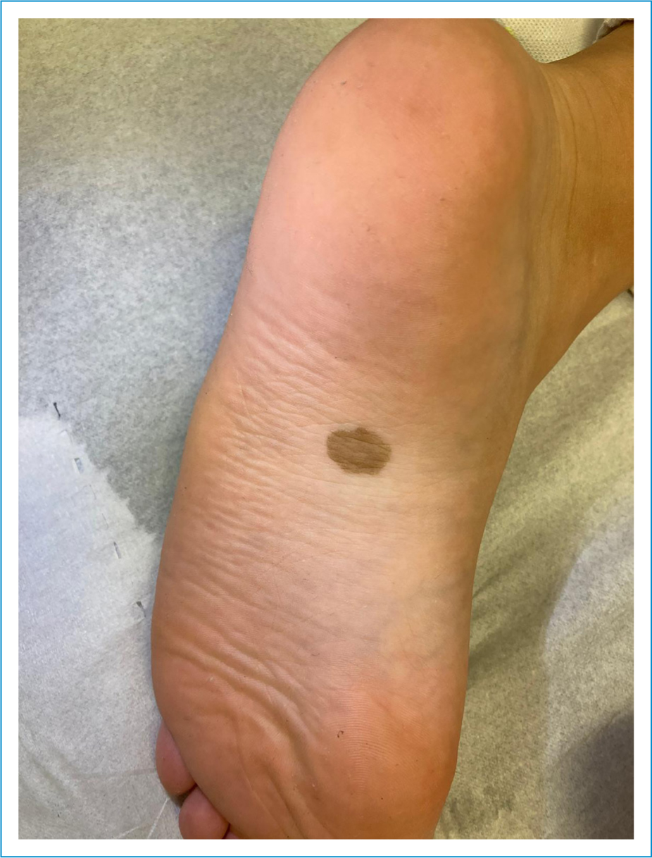
Figure 1. Light brown patch measuring about 1.5 cm, with well-defined and regular borders, and with no scaling, on the left sole.
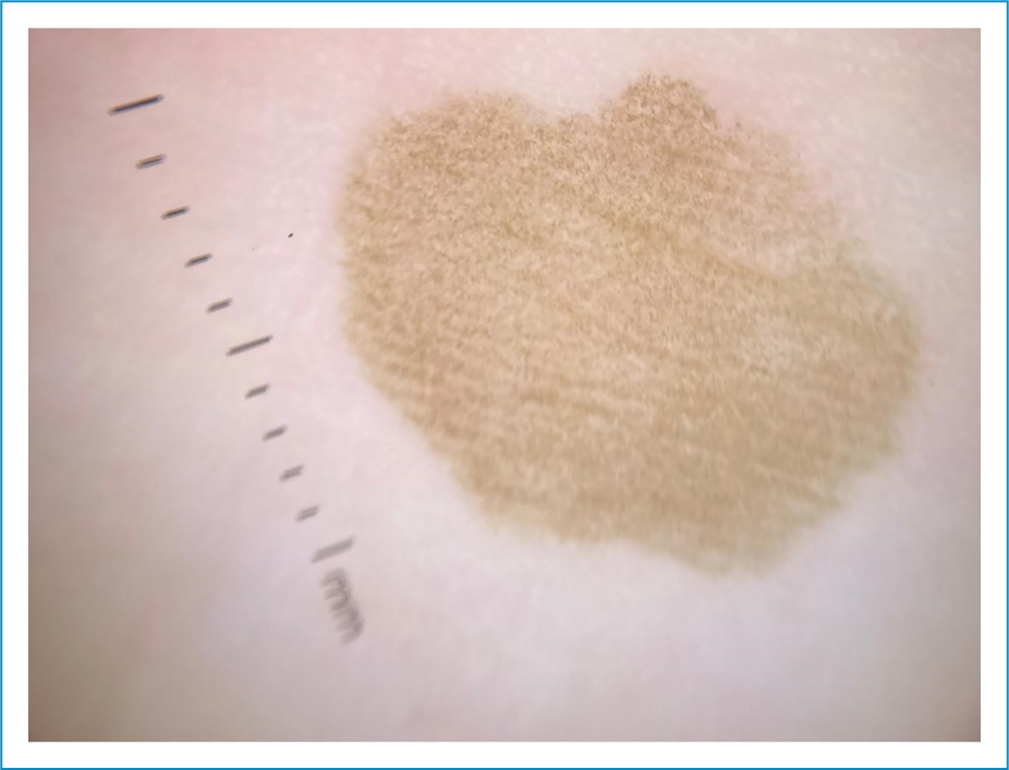
Figure 2. Dermoscopic image showing a pigmented pseudo-network with no structures suggesting a melanocytic etiology.
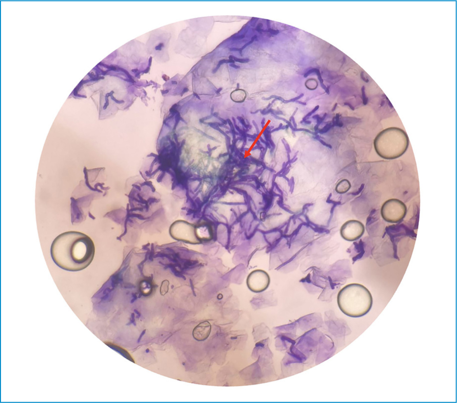
Figure 3. Tape test with methylene blue staining revealing countless hyphae.
Tinea nigra is a fungal infection caused by Hortaea werneckii,1,2 a yeast that is common in soil, and particularly prevalent in tropical and subtropical environments.1 Hyperhidrosis is a recognized risk factor.1 It is characterized by an asymptomatic brown to black macule or patch, sometimes with a slightly scaly surface, with slow, progressive growth over time.1,2 The most common sites are the palms of the hands and soles of the feet.1,2 A mycological examination is indicated upon clinical suspicion.1 At the patient’s bedside, dermoscopy and fresh examination using the tape test with methylene blue staining are important diagnostic aids.1,2 Pigmented spicules outlining a reticular pattern are characteristic dermoscopic findings, helping to differentiate it from other pigmented lesions, particularly melanocytic lesions.2 In direct examination, the fungus can be observed in its filamentous form, with multiple hyphae.1 Treatment with a topical imidazole antifungal for two to four weeks is generally sufficient.1,2
Author contributions
M. Mendes de Carvalho: contributed to the writing of the manuscript, performed the literature review, and was involved in the collection of images for the manuscript. C. Amaro: contributed to the critical revision of the manuscript, approved the final version, and participated in the collection of images. G. Catorze: contributed to the critical revision of the manuscript and assisted in the collection of images.
Funding
None.
Conflicts of interest
None.
Ethical considerations
Protection of humans and animals. The authors declare that no experiments involving humans or animals were conducted for this research.
Confidentiality, informed consent, and ethical approval. The authors have followed their institution’s confidentiality protocols, obtained informed consent from patients, and received approval from the Ethics Committee. The SAGER guidelines were followed according to the nature of the study.
Declaration on the use of artificial intelligence. The authors declare that no generative artificial intelligence was used in the writing of this manuscript.
