

Keypoints
What is known
– Clinicians should consider a GNAO1-related disorder in patients with global developmental delay, hypotonia, epilepsy and/or movement disorder.
– GNAO1-related disorder has a considerable phenotypic spectrum, in relation to the specific pathogenic variants in the GNAO1 gene.
– Affected families should receive reproductive genetic counselling to understand the implications for future pregnancies, including the potential presence of gonadal mosaicism, which could affect the transmission of genetic variant.
What is added
– Next-generation sequencing technologies offer hope in the diagnosis to many patients with cryptogenic encephal-opathies.
– The c.607G>A variant in GNAO1-related disorders is linked to a severe phenotype of global developmental delay, hypotonia, epilepsy and movement disorder.
– Early recognition and diagnosis support informed decision-making, help to manage parental expectations and guide supportive care.
Introduction
Reported for the first time in 2013, GNAO1-related disorder or GNAO1-associated epileptic encephalopathy and movement disorder, is a rare neurodevelopmental group of disorders that manifests in childhood or, more rarely, in early adulthood and is caused by autosomal dominant variant in the GNAO1 gene.1,2 The majority of cases are associated with de novo autosomal variant although gonadal mosaicism has also been reported.3,4
The GNAO1 gene encodes the α subunit of the guanine nucleotide-binding protein (Gαo), a heterotrimeric G protein family, together with a β and γ subunit. This protein plays a crucial role in signal transduction and is essential for G protein-coupled receptor (GPCR) function.1 The Gαo subunit is responsible for binding guanine nucleotides (GDP and GTP) and interacting with GPCRs to mediate cellular signaling.5 Gαo therefore participates in regulating neuronal excitability and neurotransmission and has been shown to be essential for nervous system development and functionality.6,7,8
Different molecular mechanisms have been proposed to explain GNAO1-related disorders. A well-substantiated model proposes that the majority of variants show loss of function in the transmission of GPCR signals due to distinct mechanisms that affect the G protein cycle, including impaired downstream protein binding and signaling. Additionally, some variants have also shown dominant-negative effects that interfere with the function of normal Gαo. These are two identified mechanisms responsible for the disruption of GPCR signaling.9
GNAO1 variant were first reported in association with early-onset epileptic encephalopathy syndromes.5 Since the first reports, a series of patients have been reported to have severe movement disorder as a prominent manifestation in association with developmental delay.10 GNAO1-related disorder is thus responsible for multiple neurologic manifestations of varying severity, including neurodevelopmental delay, movement disorder and/or epilepsy.
One of the most severe variants described to date is c.607G>A, producing the Gαo [G203R] protein variant.11 As of March 2025, 23 cases with this variant had been described worldwide.1,10,12–24 Reported cases include early-onset epileptic seizures, severe movement disorder, developmental and intellectual delay as well as brain malformations (Table 1). Other variants (i.e., c.644G>A) seem to produce much milder clinical outcomes, mostly late-onset hyperkinetic movement disorder.11
Table 1. GNAO1-related disorder: p.Gly203Arg (c.607G>A)-phenotype correlations
| Authors | Number of patients | Clinical characteristics | ||
|---|---|---|---|---|
| Epilepsy | Movement disorder | Developmental delay | ||
| Nakamura et al. (2013)1 | 1 | Focal seizure (tonic upgaze), tonic seizure at 5 years. Intractable (several times per day) | Opisthotonic posture, severe chorea, athetosis | Developmental delay at 7 months |
| Saitsu et al. (2016)10 | 1 | Tonic-clonic seizures at 7 days. Complex partial seizures. Intractable seizures | Severe chorea | Intellectual disability, motor developmental delay |
| Thiel et al. (2023)13 | 6 | Focal and generalised seizures | Focal, multifocal and generalised dystonia, paroxysmal chorea, hyperkinetic crisis, myoclonus | No milestones reached |
| Arya et al. (2017)14 | 1 | Generalized-onset seizures | Severe chorea | Severe global developmental delay |
| Schorling et al. (2017)15 | 2 | Different types of seizures: spasm like tonic seizures with turning to either side | Opisthotonus as well as episodes of dyskinetic movements | Severe intellectual disability and motor developmental delay |
| Xiong et al. (2018)16 | 1* | Focal motor seizures | Spasm and tonic spasm | Severe global developmental delay |
| Graziola et al. (2021)17 | 1 | Focal seizures | Generalized dystonia | Significant developmental delay, with severe language impairment and poor motor development |
| Lee et al. (2021)18 | 1 | Unknown | Intractable dystonia | Profound psychomotor retardation |
| Yang et al. (2021)19 | 1† | Focal seizures | Dystonia | Global developmental delay |
| Krygier et al. (2022)20 | 1 | Bilateral focal seizures. Reflex seizures in the form of epileptic spasms with flexing of the trunk, loss of awareness, and generalized stiffness | Generalized chorea | Global developmental delay |
| Liu et al. (2022)21 | 1 | Focal seizures, epileptic spasm | No movement disorder described | Global developmental delay |
| Domínguez-Carral et al. (2023)22 | 3 | 1: unknown form of epilepsy in neonatal period 2: daily seizures 3: no epilepsy |
All with choreodystonia | Global developmental delay (all 3 with no head control; 1: absent language development, 2: babbling, 3: guttural sounds) |
| Gambardella et al. (2023)23 | 2 | Focal seizures | Chorea and dystonia | Severe global developmental delay |
| Li et al. (2023)24 | 1‡ | Focal seizures, generalized tonic clonic seizure, status epilepticus | Chorea and dystonia | Severe global developmental delay |
*Deceased at the age of 12 months due to severe pneumonia.
†Deceased at the age of 10 months due to milk choking and asphyxia.
‡Deceased at the age of 4 years due to exacerbation of dystonia that was triggered by infection, lasting for hours to days, ending up in dystonic state with rising creatine kinase and renal failure causing multiple organ dysfunction and death.
In patients with GNAO1 variant and epilepsy, no specific electroencephalogram (EEG) pattern has been described; rather, features are concordant with an epilepsy phenotype (for example, hypsarrhythmia in cases of infantile spasms or focal epileptiform discharges in focal seizures).25 Seizures can be controlled to some degree with anti-seizure medications (ASM), although multiple drugs are often required. A 100% seizure reduction has been reported with perampanel in a small case series, thus supporting its administration in these patients. These findings warrant further investigation into the mechanism of GNAO1 and glutamate transmission to explain the therapeutic results with perampanel.26
Among patients with movement disorders, tetrabenazine has been appointed the most effective drug.6 Responses have also been reported with trihexyphenidyl, topiramate and levetiracetam, the latter participating in the suppression of both movement disorders and epilepsy. In drug refractory movement disorders, deep brain stimulation targeting the globus pallidus appears to be an effective treatment despite there not being substantial data on long-term sustained efficacy.27,13 Interestingly, dietary zinc salt supplementation in a Drosophila model of GNAO1-related disorder demonstrated motor function restoration and longevity of mutant flies, revealing a potential therapy for patients with one of the three variants studied (Gly203Arg, Glu246Lys and Arg209Cys).28
Despite available therapies showing some efficacy in controlling epilepsy and movements disorders, no drug seems to be able to mitigate developmental delay.15
Case report
We report the case of GNAO1-related disorder, diagnosed in a Portuguese patient in 2021, caused by the c.607G>A variant, producing the Gαo [G203R] protein variant.
We report the case of a female infant with non-consanguineous healthy parents, born after an uneventful gestational period, except for a history of insulin-treated gestational diabetes and oligohydramnios. Pregnancy screenings were normal and the child was born at 37 weeks and 6 days, with an APGAR score of 8/10 and appropriate somatometry at birth.
The infant revealed appropriate development and unremarkable health until the age of two months, when she was taken to the Pediatric Emergency Department presenting with generalized tonic seizures with fixed gaze and facial flushing, followed by a cyclical screaming cry lasting two to three minutes, and focal aware clonic seizures of the right upper limb followed by a cyclical screaming cry lasting two minutes. Characteristic crying was observed in the Emergency Department (Video 1). On admission, the infant was apyretic and the general examination was unremarkable.
An interictal EEG revealed bilateral centrotemporal paroxysmal activity, which was more intense in the right-hand hemisphere (Fig. 1). A brain magnetic resonance (MR) with spectroscopy was unremarkable. The cerebrospinal fluid composition and pressure were within reference ranges. Blood tests were normal, including blood gas tests. Empirical therapy with cefotaxime, vancomycin and acyclovir was given, and levetiracetam was initiated. While receiving levetiracetam, the patient developed episodes of right-sided eyelid myoclonia together with focal aware clonic seizures of the right upper limb (Video 2), prompting the initiation of phenobarbital. A trial with pyridoxine, pyridoxal-5-phosphate and folinic acid was also initiated, with no clinical or EEG improvement.
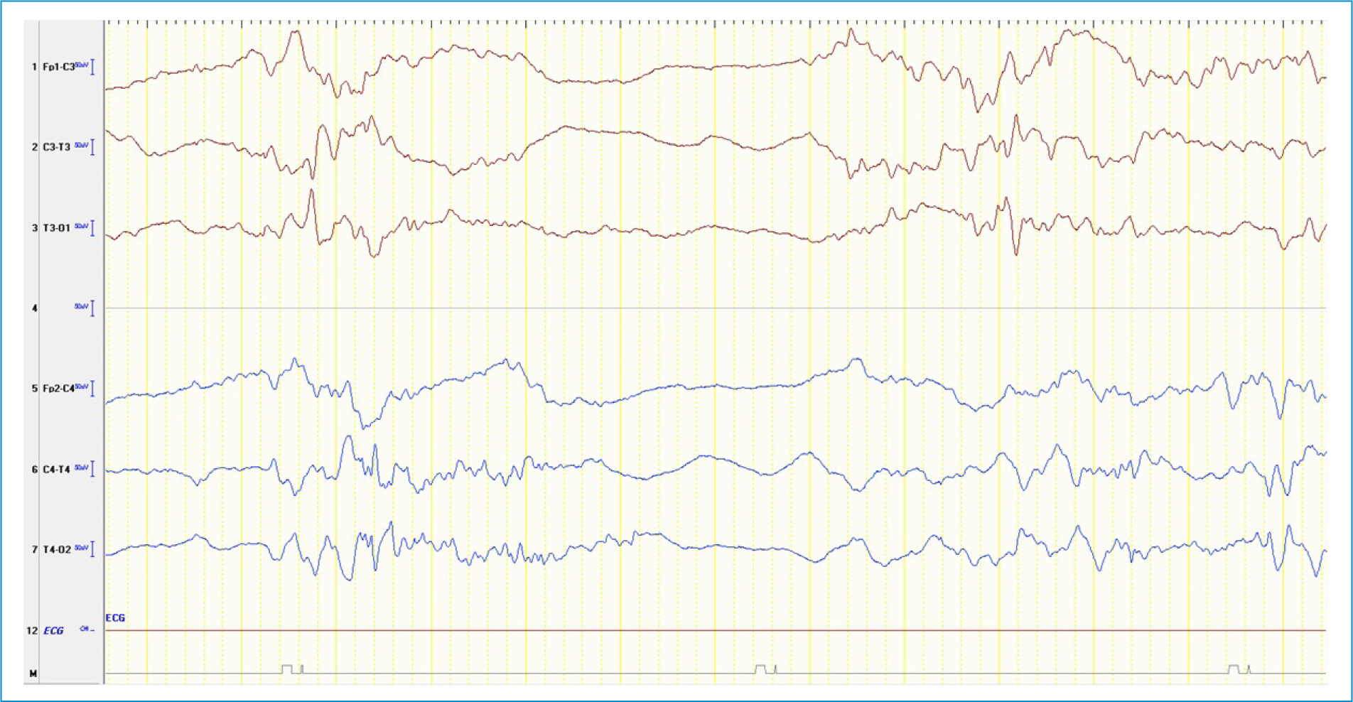
Figure 1. Initial electroencephalogram. Bilateral centrotemporal paroxysmal activity, which was more intense in the right-hand hemisphere, and sporadic broad frontal spikes. There are some episodes of flattening of baseline electrogenesis, lasting two to four seconds, but with no criteria for a burst-suppression pattern.
Further etiologic testing, including serum screening for herpes family viruses and blood cultures, were all negative. Thyroid function was normal. The neonatal screening program, via blood spot tandem mass spectrometry, was negative. Further investigation of metabolic defects, with testing of urine organic acid, free total carnitine and plasma acylcarnitine profiles, was normal.
After starting levetiracetam (40 mg/kg/day) and phenobarbital (5 mg/kg/day), there was no recurrence of seizures and the child maintained good vitality, spontaneous and symmetrical motor activity and adequate muscle tone and reflexes. The patient was discharged after 17 days and referred to a neuropediatric consultation.
Given the unclear etiology after the initial workup, a targeted gene panel sequencing for epilepsy (WES-based NGS panel for 596 genes, including CNV analysis) was requested and revealed a heterozygous pathogenic variant (c.607G>A p.(Gly203Arg)) in gene GNAO1, confirming a genetic cause for the patient’s manifestations. The parents’ genetic screening for the variant was negative, suggesting a de novo mutation in our patient.
At five months of age, developmental delay ensued. It initially manifested with poor cephalic control and rapidly progressed to global hypotonia. At eight months of age, more severe motor dysfunction was present with frequent chorea-like movements, evolving to frequent dystonic opisthotonus episodes at twelve months of age, with only a partial response to pharmacotherapy with trihexyphenidyl. These involuntary movements were responsible for complicated feedings and led to frequent choking, leading to a gastrostomy at the age of 24 months. The patient was able to babble until she was one year old. However, she never advanced further in language. Constipation became a persistent issue at nine months of age, with the need for daily therapy with laxatives. Maintenance insomnia also became an issue, with the need for pharmacotherapy at bedtime.
The patient is currently aged three and a half and is medicated with clonazepam (1.2 mg/day, 0.1 mg/kg/day, at night), trihexyphenidyl (9 mg/day, three times/day) and tetrabenazine (10 mg three times/day, 3 mg/kg/day) for treatment of abnormal movements. She has a paucity of movements and mild dyskinesia despite medication. Epilepsy control has been challenging despite the use of five anti-epileptic drugs (levetiracetam 50 mg/kg/day, twice/day; topiramate 10 mg/kg/day, twice/day; oxcarbazepine 30 mg/kg/day, twice/day; gabapentin 100 mg, three times/day and perampanel 4 mg/day, at night). Neurogenic bladder with detrusor sphincter dyssynergia and bladder wall thickening, motivated long-term catheterization and therapy with diazepam (0.4 mg/kg/day, three times/day) and oxybutynin (0.4 mg/kg/day, twice/day).
The child currently maintains generalized tonic seizures of all four limbs, associated with nystagmus and guttural noises, lasting around one minute, with eight to 10 episodes per day. She has very poor interaction and functional eye contact and does not communicate verbally. The child reveals oral and limb dyskinesias, a significant paucity of movements and is globally hypotonic. Feeding via a gastrostomy button has allowed nutrition, with ponderal evolution in the third percentile and length in the 15th percentile (according to WHO growth curves). Multiple respiratory infections have motivated six hospital admissions in the most recent year, with progressive gain of resistance to antibiotics and chronic colonization to Pseudomonas aeruginosa. Multiple periods of inconsolable crying and irritability justified starting treatment with buprenorphine (currently with 17.5 mcg/dose, 72/72 h), allowing for a better control of understandable discomfort.
Discussion
This patient’s age at onset and clinical manifestations were consistent with a form of early-onset epileptic encephalopathy. These syndromes result from identifiable primary causes, such as structural, neurodegenerative, metabolic, or genetic defects.30 Next-generation sequencing technology has revolutionized our ability to sequence DNA at the whole exome level and numerous studies in epilepsy genetics have successfully identified an increasing number of genetic causes.31 The number of cases with GNAO1 variant is expected to rise thanks to an increasing number of variants identified and as further sequencing of patients with cryptogenic encephalopathies is performed.32
Complete pathophysiologic mechanisms underlying this disorder are yet to be elucidated, but variant in GNAO1 and other G-protein subunits in early-onset movement disorders support the notion that disruption of the G protein GPCR pathway axis is a key contributor to the pathophysiology of movement disorder.6 Impaired modulation or transduction of transmembrane signaling, presynaptic autoinhibitory effects and altered neuronal excitability are all putative disease mechanisms that might explain the co-occurrence of multiple neurologic manifestations such as epilepsy and movement disorder.6Despite distinct behavioral and clinical manifestations between different GNAO1 variants, the widespread occurrence of neurodevelopmental abnormalities suggests a shared defect among most GNAO1 variants. At later stages, the different variants deviate from each other into different phenotypes, suggesting that the Gαo-controlled biochemical/physiological processes continue to be affected differently for each variant postnatally.11
Previously published case reports of the c.607G>A variant describe early-onset, sometimes days after birth, epileptic seizures, motor dysfunction, developmental delay and intellectual disability. The case we report has a similar phenotype to those previously described. As seen in this patient, the initial brain MR is usually normal, but delayed myelination, thin corpus callosum and progressive cerebral, cerebellar and brainstem atrophy may develop at later stages.3,6
The current incomplete understanding of the underlying pathophysiology has made treatment strategies hard to develop. Since no curative treatments are available, treatment is aimed at relieving individual symptoms, mostly with mild and temporary relief, as observed in this patient.
Timely identification allows healthcare providers to tailor interventions that address specific symptoms and improve the child’s quality of life. Additionally, it enables families to access appropriate resources, genetic counseling and potential clinical trials.
Author contributions
P. Sousa: conceptualization; K. Gouveia de Freitas, F. Caldeira, A. Forno: data collection and analysis; K. Gouveia de Freitas, A. Forno: drafting of the manuscript; P. Sousa, A. Forno, F. Caldeira: critical review of the manuscript; A. Forno and P. Sousa: final approval of the article.
Funding
None.
Conflicts of interest
None.
Ethical considerations
Protection of humans and animals. The authors declare that no experiments involving humans or animals were conducted for this research.
Confidentiality, informed consent, and ethical approval. The authors have followed their institution’s confidentiality protocols, obtained informed consent from patients, and received approval from the Ethics Committee. The SAGER guidelines were followed according to the nature of the study.
Declaration on the use of artificial intelligence. The authors declare that no generative artificial intelligence was used in the writing of this manuscript.
Data availability
The data that support the findings of this study are available from the corresponding author upon reasonable request.
