

Keypoints
What is known
– Pulmonary calcifications can be of infectious, neoplastic or environmental etiology, and are rare in the pediatric age.
– In rare cases, unresolved calcification may, in time, culminate in the ossification of the pulmonary parenchyma.
– Tuberculosis is a well-known cause of pulmonary calcifications, mainly dystrophic calcifications in the inactive phase of the disease.
What is added
– In extreme cases, such as this one, the ossification process can involve the entire lung parenchyma.
– Differential diagnosis is broad and challenging, and includes tuberculosis as well as other infectious and non-infectious diseases.
– In cases where there is complete functional amputation of the lung, pneumonectomy can offer an improvement in lung function.
A previously healthy seven-year-old boy from Guinea-Bissau was transferred to Portugal to investigate a long-term pulmonary disease. Symptoms began two years prior with a persistent cough. A year later, he was diagnosed with left-sided pleural effusion, which was drained and showed purulent characteristics. Multiple courses of antibiotics were prescribed, with little to no response, and the chest tube scar did not heal properly.
On admission, the patient appeared malnourished but had no respiratory distress or hypoxemia. Persistent seropurulent drainage from the scar and decreased breath sounds over the left lung field were found. Computerized tomography showed multiple scattered calcifications and consolidation of the entire left lung (Figs. 1 and 2). Pulmonary scintigraphy confirmed functional amputation of the left lung. Cultures from sputum, blood and wound exudate were negative. A tuberculin skin test (TST) and interferon-gamma release assay (IGRA) were both positive.
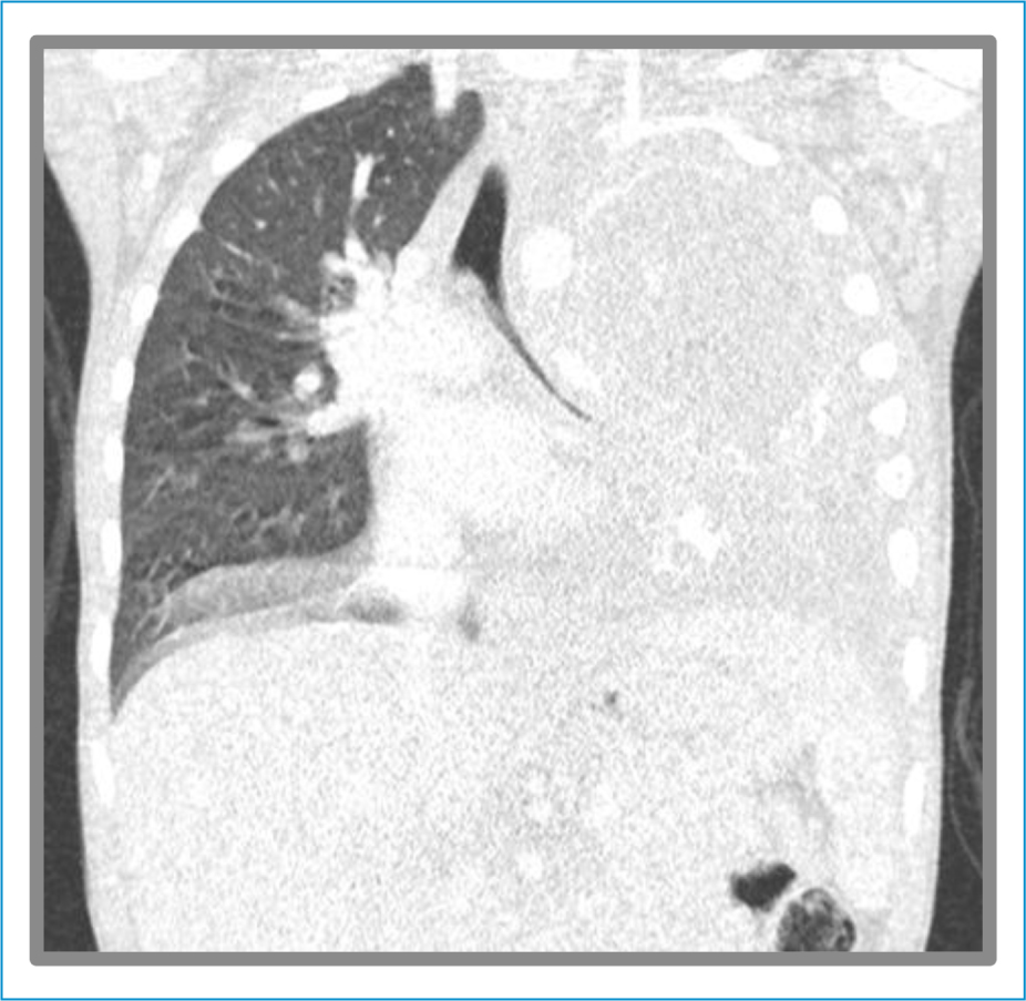
Figure 1. CT images showing complete ossification of the left pulmonary parenchyma. Coronal plane.
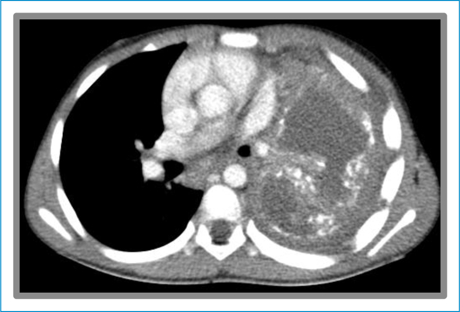
Figure 2. CT images showing complete ossification of the left pulmonary parenchyma. Transverse plane.
Quadruple antimicrobial therapy was started, leading to healing of the scar. Due to irreversible pulmonary damage with impaired lung function, a pneumonectomy was performed. The histopathological report (Fig. 3) revealed multiple calcifications, several areas of osseous metaplasia and pulmonary cavitation. No microorganisms or neoplastic tissue were identified. He completed eleven months of antimicrobial therapy with a positive clinical outcome. Post-operative lung function showed improvement compared to the pre-operative assessment, particularly regarding the forced vital capacity score.
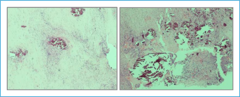
Figure 3. Histology of the operatory piece of the lung, showing complete ossification of the lung parenchyma. Hematoxylin and eosin coloration. Magnification 400x.
Pulmonary calcifications are mainly caused by two mechanisms: the metastatic form is a disseminated process that affects both the lungs and other organs;1,2 dystrophic calcifications are localized processes that follow a pulmonary insult, as in this case.1,2 In rare occasions, unresolved insults culminate in ossification of the pulmonary parenchyma.1–3
Multiple etiologies were considered for this case. The absence of histopathological neoplastic tissue rules out this hypothesis. The response to antimicrobial therapy along with the positive IGRA and TST point towards tuberculosis, but are insufficient to confirm diagnosis.4 Post-traumatic or post-infectious sequelae could have also contributed to the ossification process.
Author contributions
F. Vinagre Abrantes and D. Pereira had primary responsibility for case report drafting. L. Oliveira and L. Pereira were responsable for manuscript revision and correction. All authors provided clinical care to the patient and approved the final submitted version.
Funding
None.
Conflicts of interest
None.
Ethical considerations
Protection of humans and animals. The authors declare that the procedures followed complied with the ethical standards of the responsible human experimentation committee and adhered to the World Medical Association and the Declaration of Helsinki. The procedures were approved by the institutional Ethics Committee.
Confidentiality, informed consent, and ethical approval. The authors have followed their institution’s confidentiality protocols, obtained informed consent from patients, and received approval from the Ethics Committee. The SAGER guidelines were followed according to the nature of the study.
Declaration on the use of artificial intelligence. The authors declare that no generative artificial intelligence was used in the writing of this manuscript.
